Modern technology enables scarless biopsy for early breast cancer diagnosis
Advanced three-dimensional tomosynthesis reveals tiny abnormalities through dense tissue
Breast cancer remains one of the most common types of cancer diagnosed in women across the globe, and early detection goes a long way in fighting the condition. Modern medical technology now allows for a scarless biopsy that can be utilized to safely diagnose the illness. Apollo Athenaa Women’s Cancer Centre Radiologist and Principal Lead of Women’s Imaging Jyoti Arora revealed on Friday that it is very common for women to wait far too long before seeking a professional diagnosis.
The widespread clinical delays were detailed to HT Lifestyle, which noted that women often postpone testing due to feeling no lumps, experiencing no pain, or simply being afraid of test results. These hesitation factors regularly cost lives. “The truth is that the most dangerous breast cancers are often the ones you cannot feel, and today’s technology is finding them earlier than ever before,” Arora noted.
Dangers of delaying clinical examinations
Waiting for physical symptoms to manifest increases patient risk because breast cancer does not always announce itself. Some of the most aggressive forms of the disease begin as tiny clusters of calcifications, subtle distortions in tissue architecture, or small areas of abnormal enhancement visible only on specialised imaging. By the time a lump becomes palpable, the cancer may have been quietly growing for years.
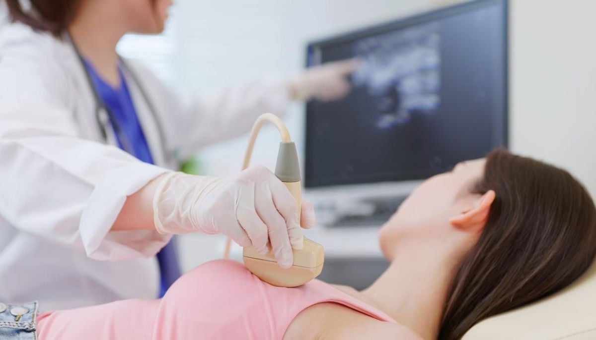
“Modern mammography, particularly 3D tomosynthesis, has transformed our ability to see through dense breast tissue,” the radiologist noted. Conventional mammography produces flat, overlapping images, but modern tomosynthesis builds a layered picture, slice by slice, to reveal previously invisible abnormalities. Cancers that medical professionals once missed entirely are now being found at the size of a grain of rice.
Advanced methods of breast cancer detection
Mammography is just one of several diagnostic tools, and it may not suffice for women with dense breasts, a strong family history, or a known genetic predisposition. Breast MRI detects hidden tumours based on their specific blood supply, making the process extraordinarily sensitive to early malignancy. After a tumour is detected, a biopsy must be performed to officially diagnose the cancer.
Earlier diagnostic methods required an invasive procedure involving general anaesthesia, stitches, permanent scarring, and anxiety-filled days of waiting. For lesions visible on an ultrasound, a standard core needle biopsy now suffices.
Arora described the updated method as “a quick, precise, minimally invasive procedure done under local anaesthetic that provides reliable tissue diagnosis without any surgery whatsoever.”
The benefits of vacuum-assisted procedures
For challenging cases where lesions are visible only on a mammography or MRI, vacuum-assisted breast biopsy, or VABB, serves as a major alternative. “VABB uses a small probe guided precisely to the target, removing multiple high-quality tissue samples with remarkable accuracy,” the radiologist stated. The advanced procedure requires no scalpel and no stitches, allowing patients to walk out of the medical facility on the same day.
Crucially, VABB is invaluable for diagnosing difficult lesions and can also be used to completely excise or remove benign non-cancerous lumps such as fibroadenomas. This technique spares women from undergoing an operation entirely. Patients are encouraged to attend regular screenings, know their breast density, and ask about an MRI if they are at high risk. “And if something is found, trust that today’s tools offer answers with minimal discomfort and potentially save your life,” Arora noted.